Lumbar Spinal Stenosis
By Julie Hastings MD and Susan Sorosky MD
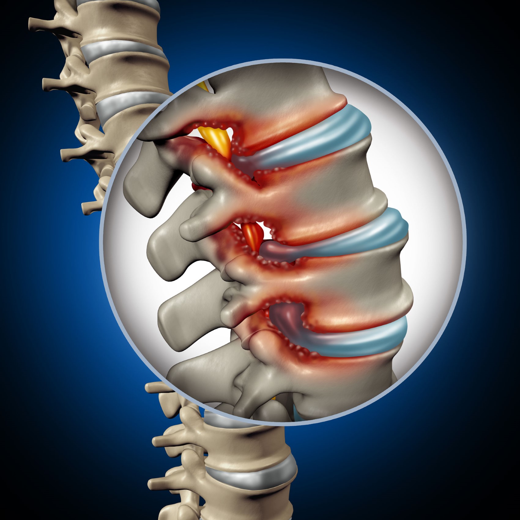
Lumbar spinal stenosis (LSS) is one of the most common pathologic conditions affecting the spine. LSS occurs when there is narrowing of the spinal canal and may involve the central canal, the lateral recess (the sides of the spinal canal) or the neural foramen (openings between the vertebrae). The spinal canal, formed from the openings within the five lumbar vertebrae, houses the spinal cord which ends in the upper lumbar region, the spinal nerves, and the spinal fluid. Each of the vertebrae are separated by intervertebral discs and connect to the vertebrae above and below through paired joints called facets. The spinal nerve roots exit from the spinal canal through openings on both sides called neural foramen and then travel to the lower extremities.
Pathology and Symptoms
LSS is most often acquired, caused by age-related degenerative changes of the spine including degenerative disc disease (narrowing of the disc), facet arthritis, osteophytes (bone spurs), spondylolisthesis (slippage of the vertebrae), and ligament thickening. Herniated discs, facet joint cysts, other spinal cysts and tumors, and spinal fractures/trauma also lead to acquired stenosis. LSS can also be congenital when a person is born with a smaller spinal canal.
Some patients with LSS are asymptomatic, while others may experience significant pain and functional impairment. The symptoms of LSS are related to where the narrowing in the spine is located, and if it is causing mechanical compression to single or multiple nerve roots. If the central canal is narrowed, multiple nerve roots may be compressed patients leading to neurogenic claudication which is the most common presenting symptom of LSS. Patients with neurogenic claudication typically report pain, heaviness, or cramping in both legs including the buttocks, thighs, and lower legs; patients may also have numbness and weakness in their legs and feet. The symptoms are usually worsened with activities that extend, or slightly arch the lumbar spine, including prolonged walking and standing. The symptoms typically improve with lumbar flexion, or bending forward as well as sitting which widens the spinal canal and relieves the mechanical pressure on the nerves. Patients often describe their pain as improving when leaning over a shopping cart or walker which is known as the “shopping cart sign.”
If the narrowing involves the lateral recess or foramen, patients may experience radicular pain (“sciatica”) that radiates along a specific dermatome (the area of sensation supplied by a spinal nerve). Patients may describe a shooting or burning type of pain, numbness, tingling, and/or weakness. Notably central, lateral recess, and foraminal stenosis can occur at the same time causing overlapping symptoms.
Diagnosis
The diagnosis of LSS is typically made by a combination of clinical evaluation and diagnostic imaging. A physiatrist is trained in taking a detailed medical and functional history and performing a comprehensive examination of the neurological and musculoskeletal systems. Imaging is also used to diagnose and assess the severity of spinal stenosis. Lumbar MRI is the most common imaging modality, followed by CT scans which are ordered for patients with non-MRI compatible pacemakers and stimulators and metal in the body, and finally CT myelograms. Electrodiagnostic studies (NCS/EMG) may also be performed by physiatrists to better characterize the severity of nerve root involvement and rule out other potential causes of leg symptoms, including peripheral nerve entrapment and peripheral neuropathy.
Treatment
Many patients with LSS experience reduced pain and improved function with conservative management. Physiatrists specialize in the design and implantation of these non-surgical plans which may include activity modification, physical therapy, epidural steroid injections, and adjunctive medications such as non-steroidal anti-inflammatories and neuropathic (nerve pain) medications. Patients with LSS often find it easier to perform aerobic exercise that promotes slight lumbar flexion, such as riding a bicycle or walking up an incline or a hill. Physical therapy protocols include flexion-based exercises focusing on core and spine stability as well as hip strengthening and spine and lower extremity flexibility exercises.
When a patient has moderate to severe pain and functional limitation, a treatment plan may also include epidural steroid injections, which interventional physiatrists are trained in performing. This type of procedure involves injecting pain relieving and anti-inflammatory medication (steroid) at the level of the lumbar stenosis under fluoroscopic (x-ray) guidance. Medical literature supports the use of these procedures to improve pain and walking tolerance, as well as to reduce opioid use and need for spine surgery. A small percentage of patients with lumbar stenosis require surgery. These patients may have intractable pain and functional limitation that has not responded to conservative management, progressive neurological deficit such as leg or foot weakness, or signs of Cauda Equina Syndrome (see below). The physiatrists at Desert Spine and Sports Physicians will help determine the best treatment plan for a patient with LSS based on symptoms, physical examination, imaging, and response to non-surgical treatments. Please contact us if you have any questions!
Please Note: If the nerves at the bottom of the spinal cord are too significantly compressed, a rare but serious diagnosis of Cauda Equina Syndrome (CES) may occur. Symptoms of CES include severe low back pain, leg weakness, sensory loss, saddle anesthesia (unable to feel anything in the body areas that sit on a saddle), bladder dysfunction (urinary retention or incontinence), bowel incontinence, and/or sexual dysfunction. This is a medical emergency, and any patient suspected of CES should report directly to the emergency room.
References:
Bagley C, MacAllister M, Dosselman L, Moreno J, Aoun SG, El Ahmadieh TY. Current concepts and recent advances in understanding and managing lumbar spine stenosis. F1000Res. 2019 Jan 31;8:F1000 Faculty Rev-137. doi: 10.12688/f1000research.16082.1. PMID: 30774933; PMCID: PMC6357993.
Bydon M, Alvi MA, Goyal A. Degenerative Lumbar Spondylolisthesis: Definition, Natural History, Conservative Management, and Surgical Treatment. Neurosurg Clin N Am. 2019 Jul;30(3):299-304. doi: 10.1016/j.nec.2019.02.003. PMID: 31078230.
Diwan S, Sayed D, Deer TR, Salomons A, Liang K. An Algorithmic Approach to Treating Lumbar Spinal Stenosis: An Evidenced-Based Approach. Pain Med. 2019 Dec 1;20(Suppl 2):S23-S31. doi: 10.1093/pm/pnz133. PMID: 31808532; PMCID: PMC7101167.
Wu L, Cruz R. Lumbar Spinal Stenosis. 2020 Dec 20. In: StatPearls [Internet]. Treasure Island (FL): StatPearls Publishing; 2021 Jan–. PMID: 30285388.
